
Figure 2.
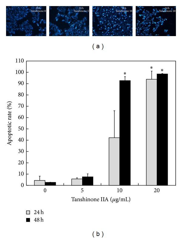
Assessment of nuclear morphological changes of KB cells exposed to Tan IIA. KB cells were treated with various contractions of Tan IIA (0, 5, 10, and 20 μg/mL) for different time periods. (a) After 24 hours, cells stained with Hoechst and exhibiting shrunken, condensed nuclei with fragmented chromosomes under fluorescent microscope were identified as apoptotic cells. (b) Apoptotic rates were calculated. The points are the mean ± SD of 2 independent experiments. ∗ indicates P < 0.05.