
Figure 1.
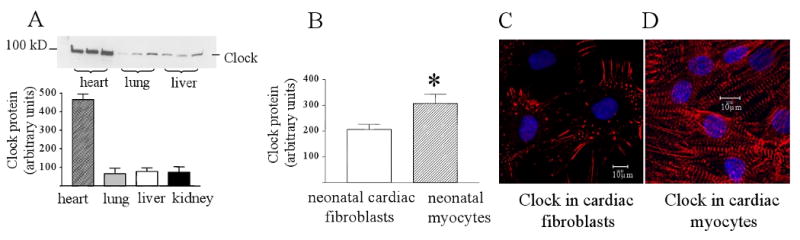
(A) Western blot of Clock protein in the adult rat heart, lung, liver and kidney. Samples were collected at Zeitgeber 9.00 hours during the resting period for rats. Clock protein expression was 5-fold higher than the three other tissues. (B) Clock protein expression in neonatal rat cardiac myocytes and cardiac fibroblasts cultured in 5% serum for 5 days. Myocytes had significantly more Clock protein than fibroblasts. P<0.05, n=4. (C) Immunostaining of Clock protein in cardiac fibroblasts. (D) Immunostaining of Clock protein in cardiac myocytes.