
Figure 2.
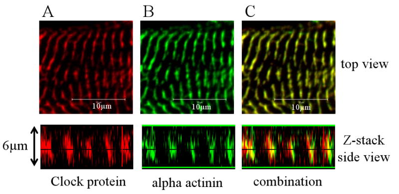
Clock localizes to the myofilaments of cardiac myocytes. (A) High resolution confocal image of Clock protein (in red) in the myofilaments of a neonatal cardiac myocyte, along with a confocal z-stack image showing that the protein is localized from the focal adhesions to the top of the cell. (B) Immunostaining for the Z-disk protein alpha actinin (in green) in the same myocyte. Panel C shows the combination of the double staining. The two proteins colocalize in the Z-disk.