
Figure 3.
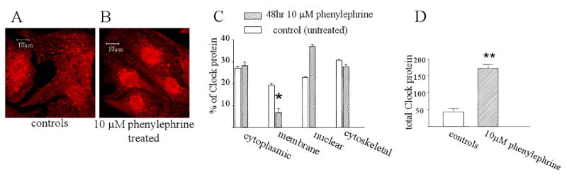
Clock subcellular localization and phenylephrine treatment. Panels A and B show immunostaining for Clock protein in untreated myocytes and those treated with 10 μM phenylephrine for 48 hours respectively. Panel C shows quantitation of Western blotting results of Clock protein distribution in subcellular fractions with and without 10μM phenylephrine treatment for 48 hours. Phenylephrine treatment results in a significant translocation of Clock from the membrane to the nuclear fraction. P<0.01, n=3 cultures. Panel D shows a Western blot of total Clock protein in untreated myocytes and following 48 hours of phenylephrine treatment. Clock protein increased about 3 fold following the drug treatment. P<0.01, n=3 cultures.