
Figure 4.
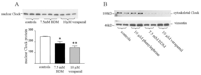
Subcellular distribution of Clock protein and myocyte cross-bridge cycling. Panel A shows a Western blot of Clock protein from nuclear fractions of untreated myocytes and those treated with either 7.5 mM BDM or 10 μM verapamil for 48 hours. There was a significant decrease in nuclear Clock in both groups. * controls vs BDM p<0.05 n=3. ** controls vs verapamil p<0.01 n=3. Panel B shows Clock protein from cytoskeletal fractions of myocytes treated with 10 μM phenylephrine, 7.5 mM BDM or 10 μM verapamil. Clock protein is present in the cytoskeleton when the myofilaments are actively contracting, whilst BDM and verapamil treatment significantly decrease its presence there.