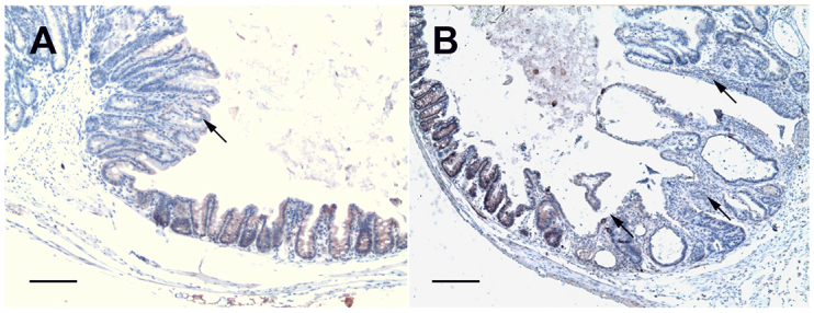
Fig. 1.

Sections of ileo-caecal regions of dexamethasone-treated SCID mice immunostained for Apc. (A) A polypoid-adenoma section from a mouse that was euthanized at 45 days post-infection showing a decrease of the intensity of cytoplasmic Apc labeling (arrow) after infection with C. parvum, whereas contiguous normal mouse tissue showed a staining pattern similar to that seen in a normal mucosa. (B) At 60 days post-infection, in a section of the ileo-caecal region taken from a mouse that had been infected with C. parvum, a decreased intensity of cytoplasmic Apc staining was observed in a polypoid-adenoma (arrow). Scale bars: 100 μm.
