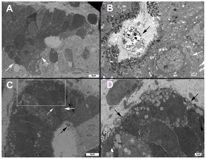
Fig. 3.

Electron micrograph of ileo-caecal regions of dexamethasone-treated mice. (A) Electron micrograph of a section of normal non-neoplastic mucosa that shows normal intercellular junctions (white arrows). (B) In SCID mice that had been infected with C. muris (black arrow), alterations in the ultrastructure of intercellular junctions (white arrow) of gastric epithelial cells were not found. (C) Dilation of intercellular spaces with extensive development of lateral membrane extensions (white arrows) was observed at the intercellular junctions of the ileo-caecal epithelia of mice infected with C. parvum (black arrow). (D) Enlarged image of the area indicate by the white box in C, which shows lateral membrane extensions (black arrows). Scale bars: 5 μm (A,C); 1 μm (B,D).
