
Fig. 2.
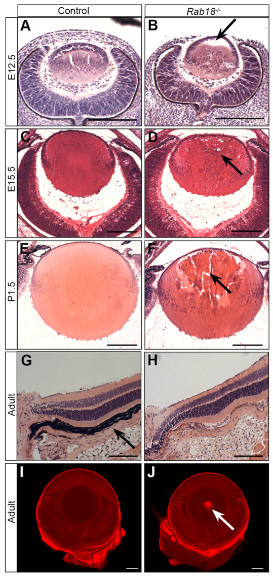
Defective pre-natal development of the lens in Rab18−/− mice. (A,B) At E12.5, Rab18−/− mice showed a delay in the filling of the lens vesicle through the delayed migration of lens fibre cells from the posterior (B, arrow), whereas wild-type littermates had a closed lens vesicle (A). By E15.5, the Rab18−/− lens vesicle had closed, but the first signs of cataract development (small vacuoles at the lens periphery) were evident (D, arrow). Signs of cataract formation were absent in controls (C). By P1.5, large vacuoles and centralised pyknotic nuclei were seen in Rab18−/− lenses (F, arrow) but not in controls (E). All other ocular structures in control (G) and Rab18−/− lenses (H) appeared normal, and no retinal degeneration was observed. Note that the Rab18−/− eye was unpigmented, owing to the origin of the mutation in 129P2-derived embryonic stem cells; consequently, the retinal pigment epithelium (arrow in G) was not easily observed. Optical projection tomography undertaken on adult wild-type (I) and Rab18−/− unpigmented eyes (J) showed a dense nuclear cataract in the centre of the Rab18−/− lens (arrow in J). Scale bars: 100 μm (A–F); 50 μm (G,H); 400 μm (I,J).