
Fig. 3.
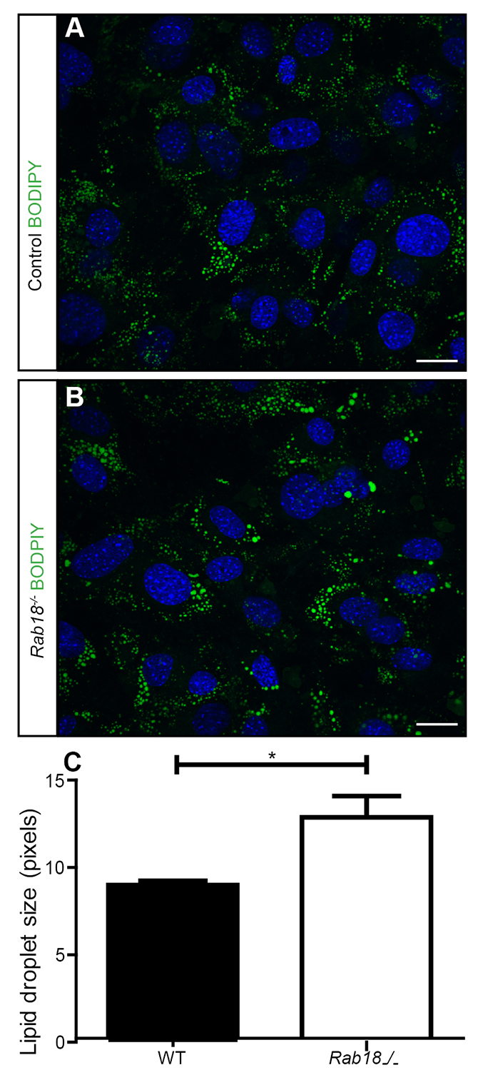
Loss of RAB18 results in the enlargement of lipid droplets in MEFs. Control (A) and Rab18−/− (B) MEFs that had been treated with oleic acid for 24 hours were fixed and stained with the neutral lipid stain BODIPY 493/503 (green), cell nuclei were stained by using DAPI (blue). (C) Quantification of lipid droplet size showed enlarged lipid droplets in Rab18−/− MEFs. n=3 mice, mean±s.e.m. (mean lipid droplet size – 8.9 pixels in control MEFs and 12.8 pixels in Rab18−/− MEFs), *P<0.05 using an unpaired Student’s t-test. Scale bars: 10 μm.