Fig. 5.
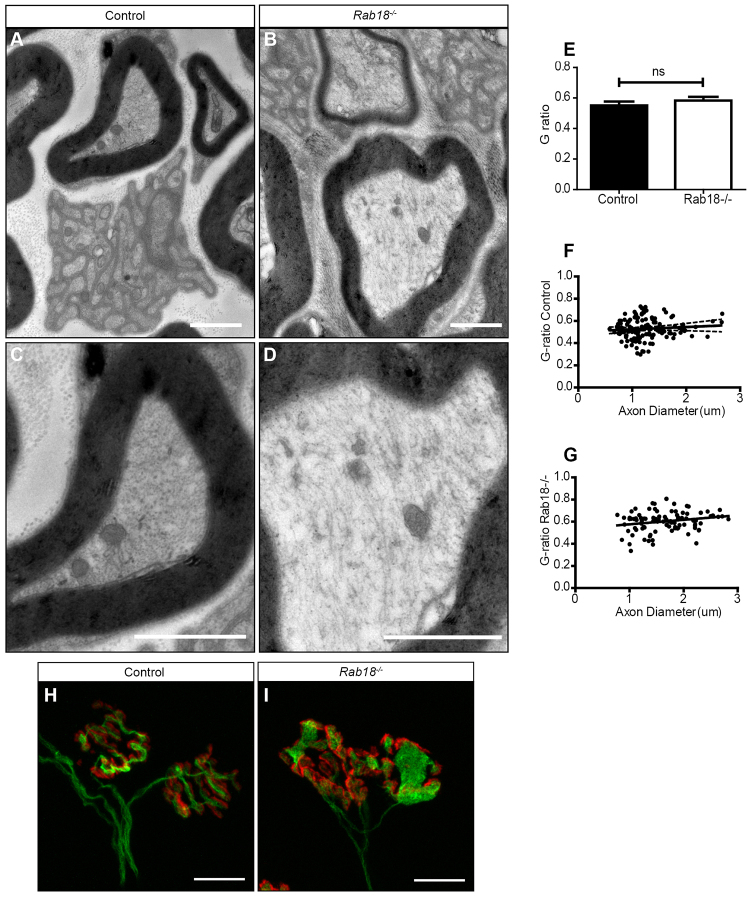
Rab18−/− mice have grossly disorganised cytoskeletons in peripheral nerves and accumulate microtubules at the neuromuscular junction. (A–D) Electron micrographs of the sciatic nerve in control (A,C) and mid-late-symptomatic Rab18−/− mice (B,D) showed normal myelination and normal Remak bundles in both genotypes. (C,D) Higher-magnification images showed disorganisation of the cytoskeleton in the sciatic nerve of Rab18−/− mice (D) compared with that of controls (C). The images are representative of that found in three animals for each genotype. (E–G) Quantification of myelination identified no abnormalities in the G-ratio (G-ratio is the diameter of an axon divided by the diameter of axon plus myelin). Statistical significance was tested by using an unpaired two-tailed Student’s t-test. G-ratio P-value=0.7610, mice (n=3) (E) or spread of axon diameter compared with the G-ratio in control (F) and Rab18−/− mice (G). (H,I) FDB muscles from wild-type (H) and mid-late-symptomatic Rab18−/− mice (I) were co-stained for β3 tubulin (green, immunostaining) and TRITC-conjugated α-bungarotoxin (red). Large accumulations of microtubules at the NMJ of Rab18−/− mice were observed. Scale bars: 2 μm (A–D); 10 μm (H,I).

