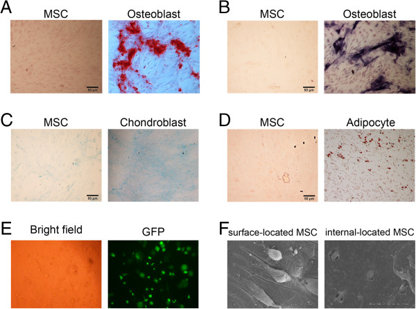
Figure 1.

Identification of multi-potential differentiation of MSCs and observation of MSCs compounded with fibrin glue. Cells were cultured and induced to differentiate into osteoblasts indicated by staining of Alizarin Red (A) and NBT-BCIP (B), chondroblasts by Alcian blue (C) and adipocytes by Oil Red O (D). (E) After fibrin glue-compounding within one week, normal cell shape under bright field and green fluorescence resulting from Ad-HGF transfection indicated the cell activity. (F) The morphologies of cells on the surface of and inside the fibrin glue were observed with the scanning electric microscopy.
