
Figure 5.
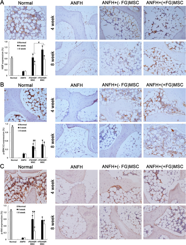
Immunohistochemical detection and semi-quantification of in vivo HGF expression, p-ERK1/2 and p-Akt. HGF expression (A), phosphorylation of ERK1/2 (p-ERK1/2) (B) and Akt (p-Akt) (C) were assayed at 4 and 8 weeks after treatment of hormone-induced ANFH with transplantation of MSCs. n = 15 or 30/group (3 sections/animal, 5 or 10 animals/group). *, P < 0.05 versus the ANFH group; #, P < 0.05 versus between the MSCs group and the MSCs + FG group (A); **, P < 0.05 versus between 4 and 8 weeks (B, C).