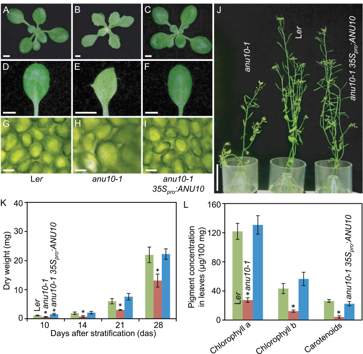
Fig. 1.
Phenotypic characterization and rescue of the anu10-1 mutant. (A–C) Rosettes, (D–F) first-node leaves, and (G–I) bright-field micrographs of the subepidermal layer of palisade mesophyll cells from (A, D, G) the Ler wild type, (B, E, H) the anu10-1 mutant, and (C, F, I) a transgenic anu10-1 35Spro:ANU10 plant. (J) Adult plants. Pictures were taken (A–I) 16 and (J) 42 das. Scale bars indicate (A–F) 2mm, (G–I) 30 μm, and (J) 5cm. (K) Dry weight and (L) pigment content in Ler, anu10-1, and transgenic anu10-1 35Spro:ANU10 plants. Error bars indicate standard deviations. Asterisks indicate values significantly different from Ler in a Mann–Whitney U-test [(K) P<0.01, n=8 and (L) P<0.05, n=4].

