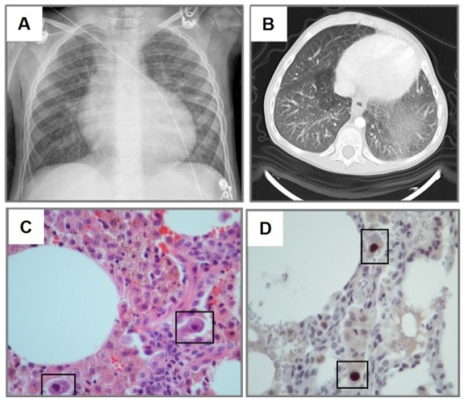
Figure 1.
Cytomegalovirus (CMV) lung infection after steroid therapy. Three year old girl treated with systemic corticosteroid for autoimmune lymphoproliferative syndrome (ALPS). Chest-X-ray (CXR) shows diffuse haziness more prominent in the left base (A), which correspond to ground-glass images in CT scan (B). H&E staining revealed monocyte infiltration with cytomegalic changes in lung biopsy (black squares in panel C). Immunohistochemical detection of CMV (brown staining in D) showed typical CMV inclusion bodies (squares). Slides shown with 100× magnification.

