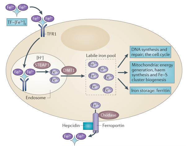
Figure 2. Key steps in mammalian cellular iron metabolism.
Iron circulates throughout the body bound to transferrin (TF), which can bind two atoms of ferric iron (Fe3+). TF-bound iron binds to transferrin receptor 1 (TFR1) on the plasma membrane of most cells, and the TF– [Fe3+]2–TFR1 complex is endocytosed. In the acidic environment of the endosome, ferric iron is released from TF and is reduced to ferrous iron (Fe2+) through the ferrireductase activity of STEAP3. The apotransferrin–TFR1 complex then recycles back to the cell surface, where apotransferrin participates in further rounds of iron uptake. In the meantime, ferrous iron is transported out of the endosome into the cytosol by divalent metal transporter 1 (DMT1), and enters the metabolically active pool of iron (the labile iron pool). Iron then traffics to multiple destinations. It is inserted into cytosolic enzymes that are required for DNA synthesis, such as ribonucleotide reductase, and is also used in haem synthesis and the biogenesis of iron–sulphur clusters, processes that occur partly in the mitochondria and partly in the cytosol. Excess iron is stored in ferritin, an iron storage protein. Iron leaves the cell through the activity of ferroportin, an iron efflux pump, and an oxidase such as ceruloplasmin or hephaestin, which can re-oxidize iron to ferric iron to enable the loading onto TF.

