Figure 4.
CAP1 Localizes to the Tonoplast and Is Expressed in Root Hairs.
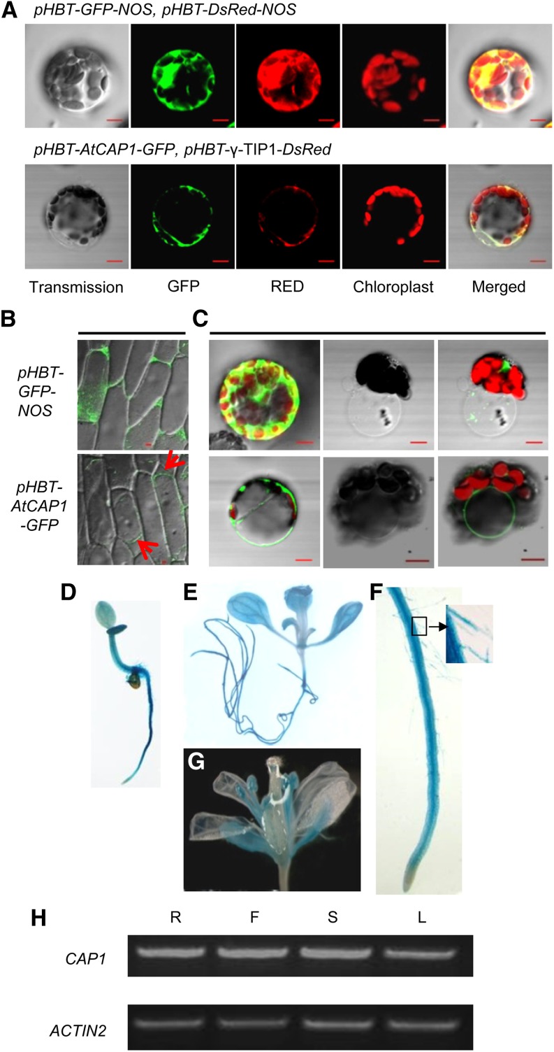
(A) to (C) Confocal analysis of the subcellular localization of CAP1. Bars = 10 µm.
(A) CAP1-fused GFP was transiently coexpressed with the vacuolar membrane marker γ-TIP1 in protoplasts. Images showed colocalization of CAP1 and γ-TIP1 (bottom panels). Empty vector pHBT-GFP-NOS expressed in protoplasts was a control (top panels).
(B) Transient expression in onion epidermal cells shows fluorescence of CAP1 in the vacuolar membrane. Arrows point to the vacuole.
(C) Fluorescence images of GFP-transformed protoplast (top panels) and GFP-fused, CAP1-transformed protoplast (bottom panels). The same protoplast before (first image) and immediately after bursting (second two images) is shown.
(D) to (G) CAP1 expression pattern. GUS staining of transgenic plants shows CAP1 expressed in roots ([D] to [F]), leaves (E), and flowers (G). Inset image shows root hairs in (F).
(H) RT-PCR indicated that CAP1 was expressed in roots (R), flowers (F), inflorescence stems (S), and young leaves (L).

