Figure 8.
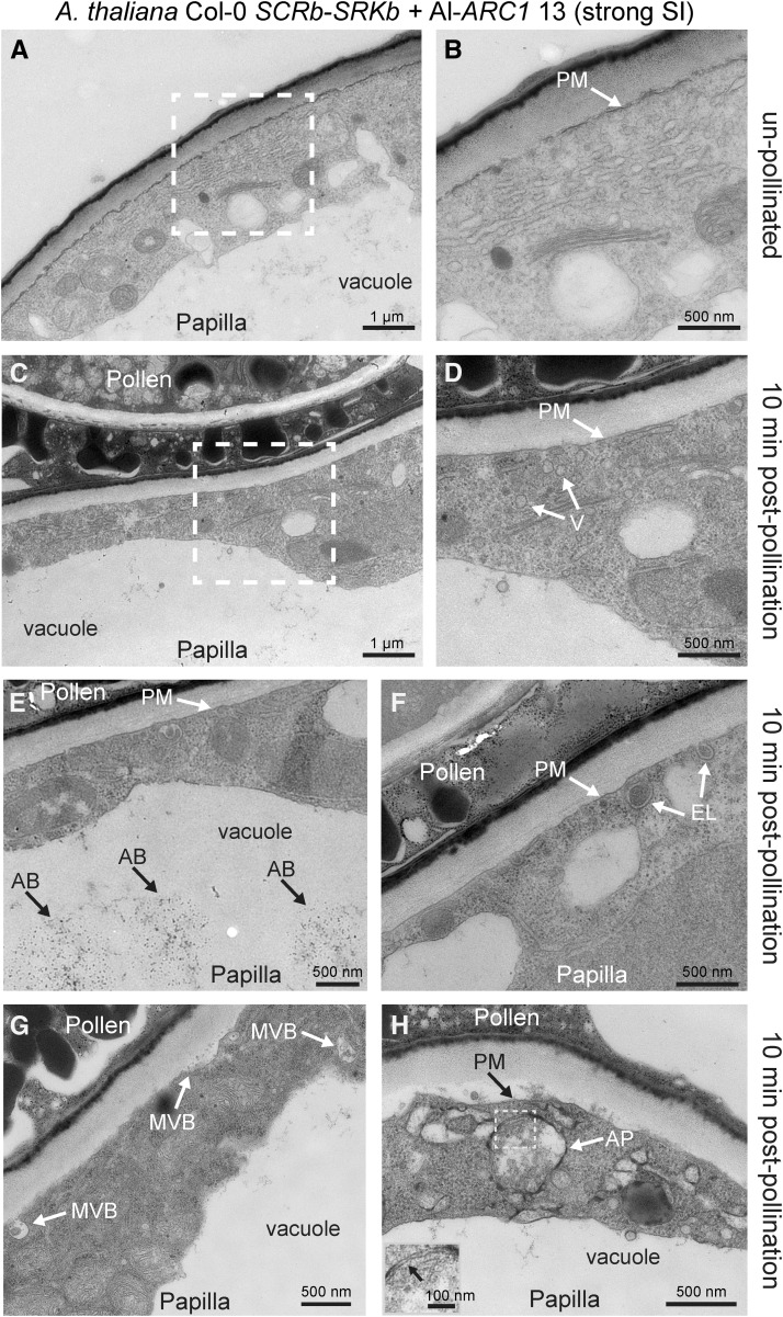
TEM Images of A. thaliana Col-0 SCRb-SRKb + Al-ARC1 Line #13 Stigmatic Papillae in Response to Self-Pollen.
(A) and (B) Unpollinated stigmatic papilla.
(C) to (H) Stigmatic papillae at 10 min postpollination. Several different structures were observed (Table 4), including vesicles (V) in the cytoplasm (D), autophagic bodies (AB) in the vacuole (E), EXPO-like (EL) structures fusing to the plasma membrane (PM) (F), and autophagosomes (A) in the cytoplasm (H). The gray boxed area in (H) shows the double membrane of the autophagosome and is displayed in the inset in the bottom left hand corner.
The white boxed areas in (A) and (C) are shown in (B) and (D), respectively. Bars = 1 µm in (A) and (C) and 500 nm in (B) and (D) to (H). Bar in (H) inset = 100 nm.

