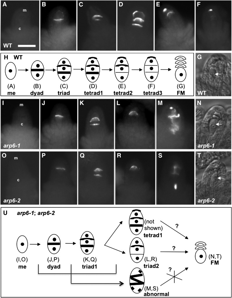
Figure 2.
Meiotic Divisions of Megasporogenesis Is Defective in arp6 Megasporocytes.
(A) to (F), (I) to (M), and (O) to (S) Epifluorescence microscopy images of aniline-blue stained wild-type, arp6-1, and arp6-2 megasporocytes, respectively.
(A) No callose staining in a wild-type megasporocyte before meiosis.
(B) Callose staining in a transverse cell plate of a wild-type dyad.
(C) Two callose bands in a wild-type triad.
(D) A megasporocyte with three callose bands in a wild-type tetrad, with the fourth band invisible in this orientation (tetrad1).
(E) A wild-type tetrad without a callose-stained wall in the chalazal end (tetrad2).
(F) A wild-type tetrad without callose-stained walls in middle and chalazal ends (tetrad3).
(G) Bright-field image of a wild-type ovule with a functional megaspore (FM; white arrow).
(H) A summary of meiosis stages in wild-type megasporocytes. Thick lines refer to callose bands.
(I) and (O) No callose staining in an arp6-1 and arp6-2 megasporocyte, respectively, before meiosis.
(J) and (P) Callose staining in transverse cell plate of an arp6-1 and arp6-2 dyad, respectively.
(K) and (Q) An arp6-1 and arp6-2 megasporocyte, respectively, with two callose bands (triad1).
(L) and (R) An arp6-1 and arp6-2 megasporocyte, respectively, with one callose band (triad2).
(M) and (S) Abnormal callose staining in an arp6-1 and arp6-2 megasporocyte, respectively.
(N) and (T) Bright-field images of arp6-1 and arp6-2 ovules, respectively, with a functional megaspore (white arrows).
(U) A summary of meiosis stages in arp6-1 and arp6-2 megasporocytes. Possibilities of a tetrad and a triad2 developing into a functional megaspore (question mark) and abnormally stained megasporocytes likely not developing into a functional megaspore (X mark). Thick lines refer to callose bands.
In each ovule, the micropylar end (m) is at the top and the chalazal end (c) is at the bottom (indicated in [A], [I], and [O]). Quantification of stages of meiotic divisions of megasporogenesis is provided in Supplemental Table 4. Bars = 20 μm.

