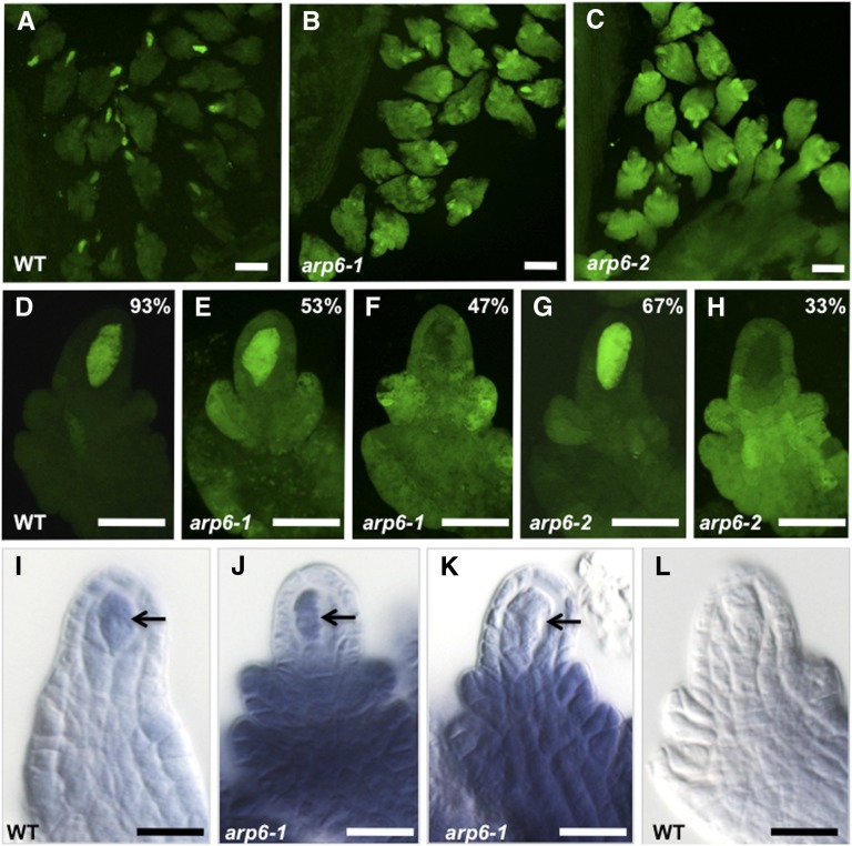
Figure 5.
DMC1 Expression in Ovules Undergoing Meiosis Is Altered in arp6 Mutants.
(A) to (H) CLSM images of pDMC1:GFP expression in wild-type ([A] and [D]), arp6-1 ([B], [E], and [F]), and arp6-2 ([C], [G], and [H]) stage 2-III/IV ovules undergoing meiosis during megasporogenesis. Images in (A) to (C) are projections of a Z stack of optical sections spanning 50 μm and were captured using identical image acquisition settings. CLSM images in (D) to (H) are 5-μm single optical sections that were captured using identical image acquisition settings. Percentages refer to number of ovules that displayed the GFP expression pattern shown in each panel; quantification details are provided in Supplemental Table 6.
(I) to (L) In situ localization of DMC1 mRNA in Arabidopsis whole-mount ovules.
(I) In situ hybridization of DMC1 antisense RNA to wild-type ovules of Col-0 ecotype (28% of the stained ovules [n = 181] showed this pattern).
(J) and (K) In situ hybridization of DMC1 antisense RNA to arp6-1 ovules.
(J) A representative arp6-1 ovule that showed DMC1 expression in the megasporocyte and nonsporogenous cells (16% of stained ovules [n = 187] showed this pattern).
(K) A representative arp6-1 ovule that showed DMC1 expression in the nonsporogenous cells but lacked DMC1 expression in the megasporocyte (72% of stained ovules [n = 187] showed this pattern).
(L) Control hybridization of wild-type ovules (Col-0 ecotype) with DMC1 sense RNA did not give any signal (100% of stained ovules [n = 47] showed this pattern).
Arrows in (I) to (K) point to the megasporocyte. Bars = 40 μm in (A) to (C), 20 μm in (D) to (H), and 10 μm in (I) to (L).

