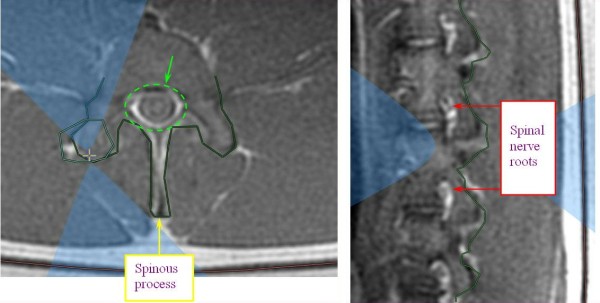
Figure 5.

MR images showing planning of sonication with focal spot on the facet joint. Appropriate beam orientation is applied in the axial and sagittal planes to protect the spinal nerve roots (white areas on the image, marked with red arrows), the spinous process (marked with yellow arrow), and the spinal canal (marked by green dashed circle) from exposure to the acoustic beam.
