Figure 8.

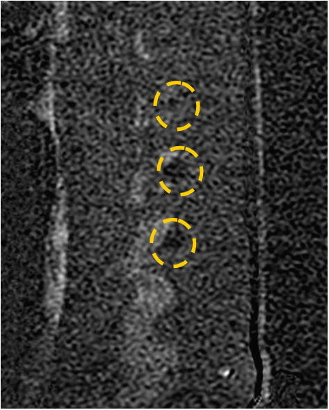
Sagittal T1-weighted contrast-enhanced subtraction MR image. The image shows NPV in the left facet joints of the treated pig that were treated with 300 J (marked by yellow circles).

Sagittal T1-weighted contrast-enhanced subtraction MR image. The image shows NPV in the left facet joints of the treated pig that were treated with 300 J (marked by yellow circles).