
Figure 2.
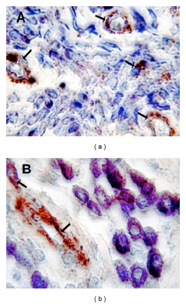
(a) Poorly differentiated G3 CMCT with high MVD. Double staining is performed combining immunohistochemistry with toluidine blue histochemistry. Many scattered degranulated blue stained mast cells. Single arrows indicate red-brown immunostained microvessels with primary anti-FVIII-RA. Note as an internal positive control the red blood cell in the lumen of microvessel. ×1000 in oil magnification. (b) Well-differentiated G1 CMCT with low MVD. Double staining is performed combining immunohistochemistry with toluidine blue histochemistry. Many scattered granulated red-blue stained mast cells. Single arrows indicate red immunostained microvessels with primary anti-FVIII-RA. Note as an internal positive control the red blood cell in the lumen of microvessel. ×1000 in oil magnification.