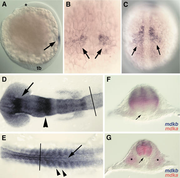
Figure 4.
Expression of mdka during embryonic development as analyzed by RNA whole-mount in situ hybridization. (A) Onset of mdka expression in paraxial mesoderm at tailbud stage (10 hpf). Anterior is to the top, dorsal to right. The anterior end of the head (asterisk) and the tailbud (tb) are indicated. Expression is restricted to mesoderm and excluded from ectodermal tissues (arrow). (B) Dorsal view at higher magnification of embryo in A, anterior is to the top. Two groups of cells (arrows) express mdka. (C) Dorsal view on embryo at 12 hpf (anterior to the top) with expanded expression of mdka in the paraxial mesoderm. Expression is excluded form the axial mesoderm. (D) Dorsal view of graded expression of mdka in the neural ectoderm of the head region at 16 hpf (14-somite stage; anterior to the left). (E) Dorsal view of posterior part of the embryo shown in D (anterior to the left). Expression of mdka in the neural tube (arrow) and somites (arrowheads) at 16 hpf. (F) Transverse section of embryos at 16 hpf at the level of caudal hindbrain (rhombomere 6; level as indicated in D). Expression of mdka (red) in the central and mdkb (blue) in the dorsal neural tube. (G) Transverse section of same embryo at the level of the spinal cord (level indicated in E). mdkb (blue) is expressed in the dorsal third of the neural tube, whereas mdka (red) is confined to the somites (asterisk) and the central neural tube, but is excluded from the floor plate.

