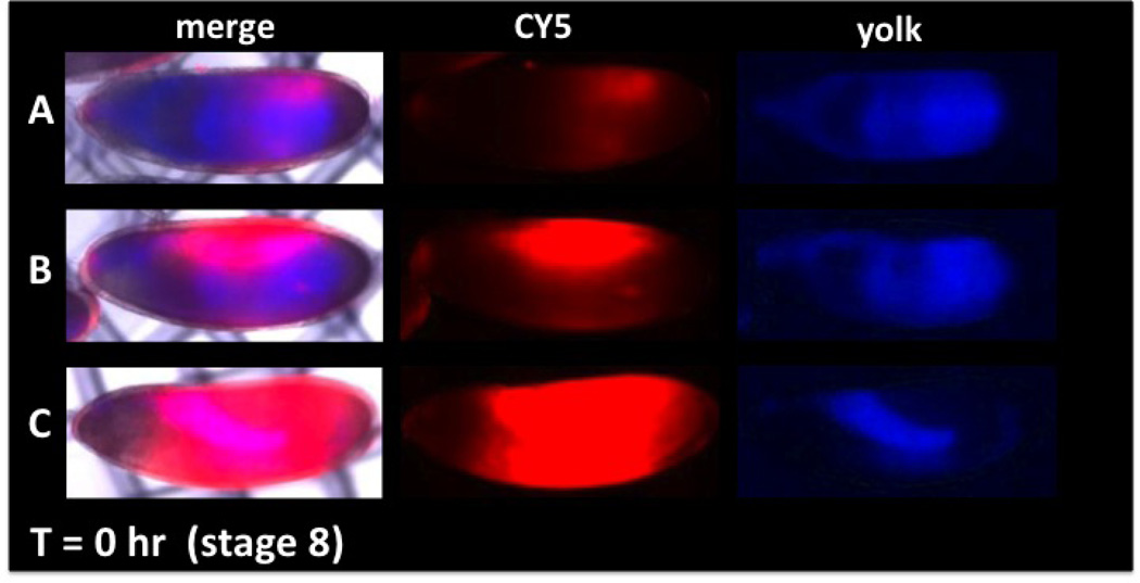
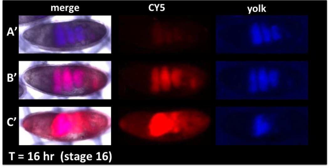
Figure 2. Permeability and viability determination with CY5 dye uptake and distribution in embryos.
(Anterior is left and dorsal is up). The degree of permeability can be assessed by CY5 uptake (red) and compared to embryo age determined by distribution of yolk (blue). A range of permeability is seen in embryos of the same stage (A, B, C, CY5). Viability can be determined by incubating embryos for a period of time (Basic Protocol 3) and observing the distribution of yolk to the involutions of the forming midgut (A’, B’, C’ yolk). Over-permeabilization (C, C’) results in decreased viability seen by the failure of midgut formation (C’, merge and yolk) and an incomplete sequestration of dye in the gut (C’ CY5). Correlation of CY5 dye uptake and viability (assessed by yolk blue autofluorescence) allows identification of optimally permeabilized viable embryos (see B and B’). Methods: A two-hour collection of embryos was aged for two hours at 18°C to obtain embryos at ~stage 8. EPS permeabilization and CY5 dye treatments were done as in Protocol 2. Embryos were placed in a notched basket with MBIM culture medium (Protocol 3) and visualized with brightfield, blue and far-red epifluorescence (A, B, C). Embryos were then aged at room temperature for 16 hours and imaged again under the same image capture settings (A’, B’, C’).


