
Figure 1.
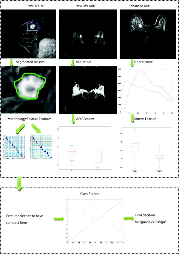
Overview of the analysis pipeline. Raw DCE-MRI is segmented to have suspicious breast mass, on which morphological and texture features are estimated. The ADC map is calculated on DWI-MRI to have the ADC feature. Kinetic curve is obtained on the enhanced image of DCE-MRI and then kinetic features are estimated. Features are extracted and selected within the combined features, and used by the classifier to predict whether the sample is malignant or benign.