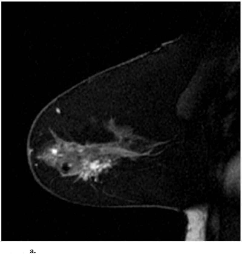
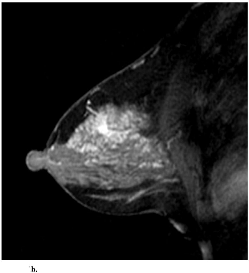
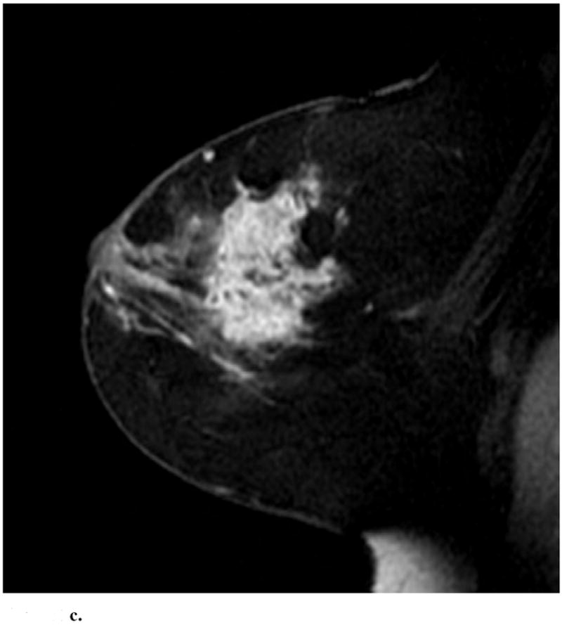
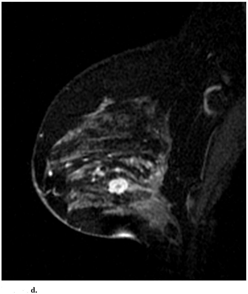
Figure 1.


Sagittal images from immediate postcontrast T1 3D-GRE sequence in 4 different patients found to have invasion in addition to DCIS. (a): Clumped ductal non-mass-like enhancement. A 2.5 cm mixed lobular and invasive ductal carcinoma was found at pathology. (b): Clumped segmental non-mass like enhancement. A 0.3 mm invasive ductal carcinoma was found at pathology. (c): Heterogeneous regional non-mass-like enhancement per Reader 1 and a lobular mass with irregular margins and heterogeneous enhancement per Reader 2. Microinvasion was found at pathology. (d): Lobular mass. Foci measuring up to 0.6 cm containing invasive lobular and ductal carcinoma were found at pathology.
