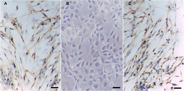
Figure 2.

Immunocytochemistry of S-100. Immunocytochemistry of S-100 in SLCs (A), BM-MSCs (B) and Schwann cells (C). After induction, the SLCs became positive in response to the S-100 antibody (brown staining in the cytoplasm), whereas the BM-MSCs were negative. Schwann cells were used as a positive control. Scale bar = 20 μm.
