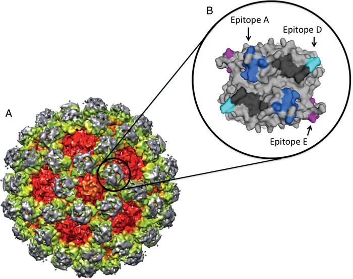
Figure 2.
GII.4 potential neutralization sites. A, A cryo electron microscopy image shows the virion structure for a GII norovirus. Approximate positions are shown for the capsid protein shell domain (red), the P1 subdomain (yellow/green), and the P2 subdomain (gray). The black circle indicates a single P2 dimer. B, A single GII.4 norovirus capsid P2 dimer (top view) is shown. Evolving surface-exposed blockade epitopes A (dark blue), D (light blue), and E (purple) are shown. Histoblood group antigen interaction sites are shown in black. These blockade epitopes represent potential vaccine and drug targets for GII.4 noroviruses.

