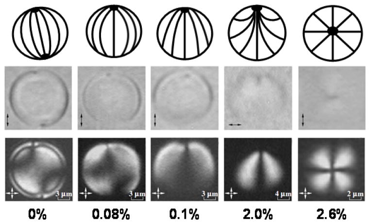
Figure 5.
Equilibrium director configurations observed in nematic LC droplets dispersed in a poly(vinyl butyral) matrix containing lecithin. The top row shows schematic illustrations of the configuration of the LC within each droplet, and the middle and bottom rows, respectively, show the corresponding bright field and polarized light micrographs of the 5CB droplets. The weight percent of lecithin doped into the polymeric matrix is indicated below each polarized light micrograph. Note that the scale-bar differs between figures. Double headed arrows in bright field micrographs indicate the orientation of the single polarizer, while double headed arrows in polarized light micrographs indicate the orientation of the crossed polarizers. Reproduced with permission.20

