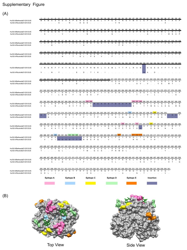
Figure 2,
Appendix. Differences in the major capsid protein (VP1) between norovirus strains GII.4 and GII.6. A) Amino acid sequence alignment of the VP1 sequences. The shell (S) domain is highlighted with a dark line, and the protruding (P) domain is highlighted with a gray line. The color code for each of the epitopes and insertions is indicated. Residue numbers are based on norovirus strain GII.4. B) Top and side views of the P domain of GII.4 norovirus showing the location of the epitopes and the carbohydrate (represented as sticks and highlighted in green) binding sites.

