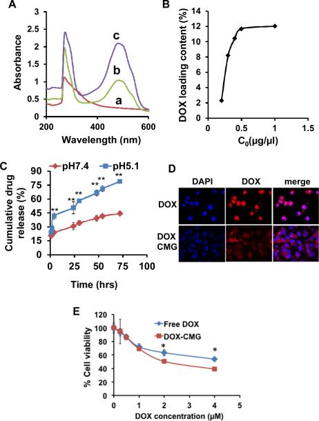
Fig.4.
(A) UV-Vis absorption spectra of (a) CMG, (b) DOX-CMG nanoparticles, and (c) DOX alone. (B) DOX loading capacity of CMG with different initial DOX concentrations. (C) Cumulative release of DOX from DOX-CMG nanoparticles at pH 7.4 and 5.1. (D) Confocal microscopic images of subcellular localization of free DOX and DOXCMG after 20 hrs incubation with A549 cells. Nuclei were stained with DAPI (**p<0.0001). (E) Viability of A549 cells treated with different concentrations of DOX and DOX-CMG (* p<0.05).

