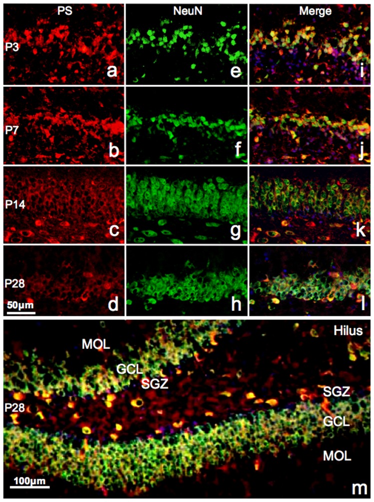
Figure 3. Fluorescent immunoreactivity of PS and NeuN.
Fluorescent micrographs of the upper blade of the dentate gyrus showing immunoreactivity of PS (red, a–d ), NeuN (green, e-h), and their merge (i–l ) at P3 (a, e, i ), P7 (b, f, j ), P14 (c, g, k), and P28 (d, h ,l ). Nuclei are stained with DAPI (blue). A low-power image of the dentate gyrus double-stained with NeuN and PS at P28 is shown in panel m. Note that the majority of prosaposin-positive neurons are also NeuN-positive at all stages.

