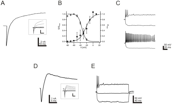
Figure 5. In vitro generated neurons display defined electrophysiological properties.
Electrophysiological properties of neurons derived from postnatal murine neurospheres. A: Exemplary voltage-gated currents elicited by a depolarizing pulse from −120 mV to +10 mV. Inserts display superpositions of consecutive current responses obtained by pulses from −120 to +30 mV in 10 mV increments. Marked transient inward sodium currents are superseded by outward potassium currents. B: Activation (filled circles) and inactivation (open circles) curves of voltage gated sodium currents. Conductance values were calculated from the respective sodium peak current amplitudes and normalized to the conductance at +10 mV. Data could be best described using a Boltzmann equation (data from 14 cells). Error bars indicate SEM. C: Firing patterns could be separated in two main groups of neurons, namely phasic (upper) and tonic (lower) patterns. D: Representative current response of a neuron, which postexperimentally proved to stain positive for biocytin and BrdU, to a depolarizing pulse from −120 to +10 mV. A prominent inward sodium current is visible at the beginning of the pulse, followed by a longer lasting potassium current. Inserts display superpositions of consecutive current responses obtained by pulses from −120 to +30 mVin 10 mV increments. E: The same neuron was capable to generate action potentials when depolarized.

