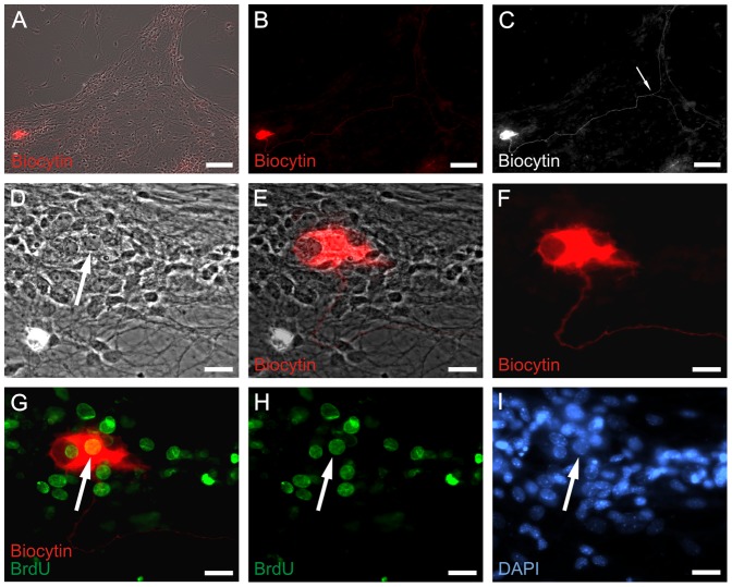
Figure 6. Electrophysiologically active neurons derive from proliferating progenitors in vitro.
Sodium channel positive neuron was derived from a proliferated ENS progenitor cell in vitro. The ENS cells were proliferated for 7 days and differentiated for 18 days prior to patch clamp recording. Under proliferation culture condition, progenitors were incubated with BrdU for 6 days. During patch clamp measurement, the recorded cell was filled with biocytin. A: Overview of combined bright-field/biocytin fluorescence (red) view of differentiated ENS cells. B: Biocytin fluorescent staining of (A). C: Enhanced black/white view of (B). The length between perikaryon and the junction of bifurcation (arrow) is 665 µm. D: Higher magnification of bright-field view shown in (A). E: Higher magnification of (A). F: Higher magnification of (B). G: Combined immunocytochemical staining of biocytin (red) and BrdU (green). H: BrdU immunofluorescence of (G). I: Nuclei staining with DAPI (blue) of the same area shown in (D–H). Arrows in (D), (G), (H) and (I) indicate the same nucleus of patched and biocytin filled neuron. Scale bars: 100 µm in (A–C); 20 µm in (D–I).

