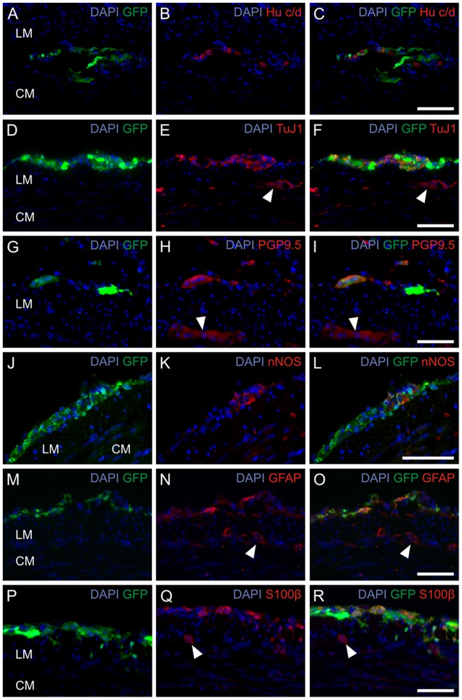
Figure 8. In vivo implanted progenitor cells differentiate into neurons and glial cells.
Immunohistochemical analysis of postnatal progenitor cells of the ENS 12 weeks after implantation into the distal colon of NOD.CB17-Prkdcscid/NcrCrl mice. Staining for the neural makers Hu c/d (A–C), TuJ1 (D–F), PGP9.5 (G–I), nNOS (J–L), GFAP (M–O) and S100β (P–R). The cells could be located within the region of the myenteric plexus (A–C) or within the longitudinal muscle layers (D–L). Ganglia of the recipient mouse are marked by white arrowheads. Nuclei were DAPI stained (blue). Scale bar: 50 µm.

