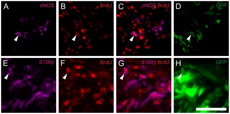
Figure 9. In vivo generated neurons derive from in vitro expanded progenitor cells.
BrdU loaded postnatal progenitor cells 3 weeks after implantation into the distal colon of NOD.CB17-Prkdcscid/NcrCrl mice. The arrows indicate triple-staining of injected cells for eGFP, BrdU and various markers as nNOS (A–D), and S100β (E–H). Triple staining provides evidence that the cells stained positive for the neural and glial markers underwent cell division and incorporated BrdU during the proliferation phase in vitro prior implantation into the mouse colon. The BrdU staining remains restricted to implanted eGFP positive cells. Cell nuclei are stained with DAPI (blue). Scale bar: 50 µm.

