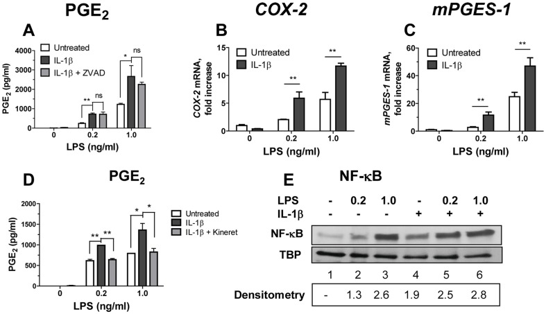
Figure 5. LPS priming is required for rIL-1β-induced COX-2 and mPGES-1 mRNA up-regulation and PGE2 production in monocytes.
(A–D) Monocytes were left untreated or were primed with LPS for 1 h (open bars) or were primed with LPS followed by addition of rIL-1β at 100 ng/ml for 18 h (solid bars). In some cultures, LPS-primed monocytes were cultured with rIL-1β in presence of ZVAD (10 µg/ml, panel A, grey bars) or Kineret (100 µg/ml, panel D, grey bars). Cell culture supernatants and cells were collected and were assayed for PGE2 (A and D), or for COX-2 and mPGES-1 mRNA (B and C), respectively. The relative values of COX-2 and mPGES-1 mRNA expression were normalized using qPCR reactions with β-actin primers performed in the same samples. The data is shown as mean PGE2±STDEV for triplicate wells in the PGE2 assay and mean mRNA fold increases over untreated cells ± STDEV calculated for triplicate wells in A and D and in B and C, respectively, **p≤0.001; *p≤0.05; ns, not significant (p≥0.05). This experiment was performed with monocytes from three separate donors with similar results. The data is shown for one representative experiment. (E) Monocytes were left untreated (lane 1) or were treated with LPS at 0.2 or at 1.0 ng/ml alone (lanes 2 and 3) or with IL-1β at 100 ng/ml alone (lane 4) or with LPS at 0.2 and at 1.0 ng/ml and IL-1β together (lane 5 and 6). At 15 min post-treatment, nuclear extracts were obtained from collected cells and resolved in SDS-PAGE. NF-κB and TBP (control) were detected by Western Blotting. Densitometry values in lower panel represent fold increase in the intensity of NF-κB band in treated over untreated monocytes in this experiment (shown in lane 1). This experiment was performed twice with the same results; data are from one representative donor.

