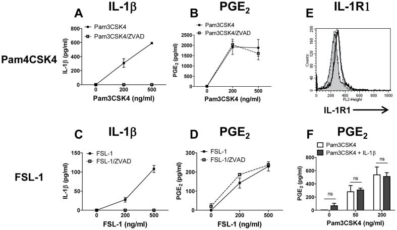
Figure 6. Caspase-1 and IL-1β do not play a role in TLR-2 ligand-induced PGE2 production in monocytes.
Monocytes were treated with Pam3CSK4 (A, B) or with FSL-1 (C, D) with or without ZVAD for 18 h (broken and solid lines respectively). Cell culture supernatants were assayed for IL-1β (A, C) and for PGE2 (B, D). (E) IL-R1 surface expression was determined by FACS analysis on untreated monocytes (dashed line) or on monocytes following activation with Pam3CSK4 or with FSL-1 at 500 ng/ml overnight (solid and dotted lines, respectively); filled histogram shows staining of untreated monocytes with isotype control. IL-1R1 ΔMFI for untreated monocytes and for monocytes activated with Pam3CSK4 or with FSL-1 was 14.2±5.1, 55.5±10.5, and 47.3±8.4, respectively, n = 3. Histograms show results from one representative experiment, each measuring at least 10,000 events. (F) TLR2 agonist does not prime monocytes for rIL-1β response. Monocytes were primed with Pam3CSK4 for 1 h at indicated concentrations (open bars) or were primed with Pam3CSK4 followed by addition of rIL-1β at 100 ng/ml for 18 h (solid bars). Cell culture supernatants were collected and assayed for PGE2 protein. The data are shown as mean ± STDEV for PGE2 and IL-1β protein concentration calculated from triplicate wells in the PGE2 assay and in IL-1β ELISA, respectively. This is representative of three experiments; ns, not significant.

