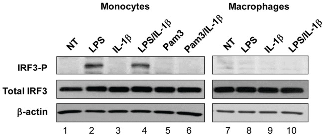
Figure 7. IRF3 is phosphorylated in monocytes but not in macrophages following activation with LPS.

Monocytes (lanes 1–6) and differentiated macrophages (lanes 7–10) were left untreated (lanes 1 and 7) or were treated for 1 h with: LPS alone at 1 ng/ml (lanes 2, 8); Pam3CSK4 alone at 50 ng/ml (lane 5); with IL-1β alone at 100 ng/ml (lanes 3, 9), or with LPS + IL1β (lanes 4, 10); or with Pam3CSK4 + IL-1β (lane 6). Following incubation, cell lysates were prepared and resolved in SDS-PAGE. Phospho-IRF3, total IRF3, and β-actin were detected after Western Blotting. The experiment was performed 3 times with similar results.
