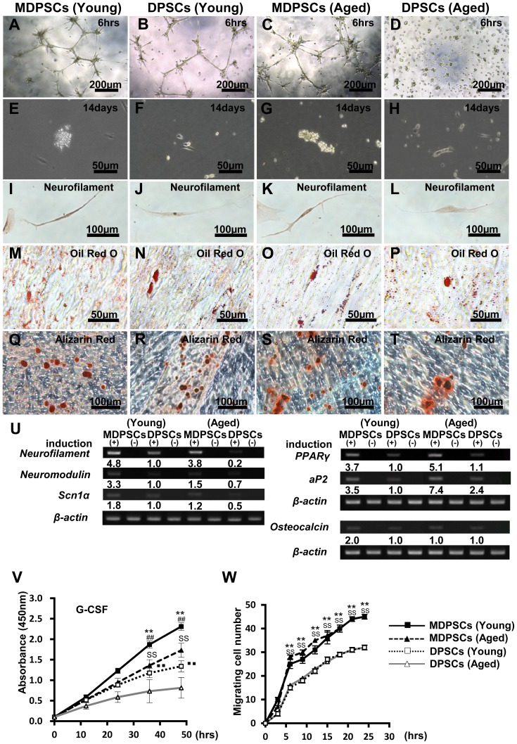
Figure 1. Multi-lineage differentiation potential and the characteristics.
The aged dental pulp stem cells (DPSCs) mobilized by granulocyte-colony stimulating factor (G-CSF) (MDPSCs) were compared with aged colony-derived DPSCs (DPSCs), young MDPSCs and young DPSCs. (A–D) The endothelial differentiation potential using the matrigel assay. (E–H) Neurosphere formation, 14 days after induction. (I–L) Neuronal differentiation potential. Fourteen days after induction of dissociated neurosphere cells. (M–P) Adipogenic differentiation potential. (Q–T) Odontoblast differentiation potential. (U) Gene expression of neurofilament, neuromodulin, and sodium channel, voltage-gated type I α (SCN1A) for neuronal markers, peroxisome proliferator-activated receptor γ (PPARγ) and adipocyte fatty acid binding protein 2 (aP2) for adipogenic markers, osteocalcin for a odonto/osteoblastic marker. (V) The proliferation analysis stimulated by 10% human serum (**p<0.01, young MDPSCs versus young DPSCs; SS p<0.01, aged MDPSCs versus aged DPSCs; ## p<0.01, young MDPSCs versus aged MDPSCs; ▪▪ p<0.01, young DPSCs versus aged DPSCs). (W) The migration analysis stimulated by G-CSF (10 ng/ml) (**p<0.01, young MDPSCs versus young DPSCs; SS p<0.01, aged MDPSCs versus aged DPSCs). Data are expressed as the means ± SD of 6 determinations. The experiments were repeated six times (6 lots), and one representative experiment is presented.

