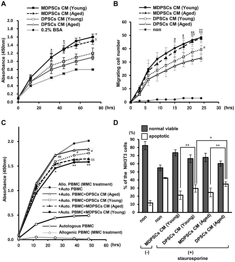
Figure 2. Effect of conditioned medium (CM) of aged MDPSCs.
Effect of conditioned medium (CM) of the aged MDPSCs compared with those of aged DPSCs, young MDPSCs and young DPSCs. (A) The proliferative effect in NIH3T3 cells. (*p<0.05, young MDPSCs CM versus young DPSCs CM; S p<0.05, aged MDPSCs CM versus aged DPSCs CM). (B) The migratory effect in NIH3T3 cells (*p<0.05, **p<0.01, young MDPSCs CM versus young DPSCs CM; S p<0.05, SS p<0.01, aged MDPSCs CM versus aged DPSCs CM; # p<0.05, young DPSCs CM versus aged DPSCs CM). (C) Mixed lymphocyte reaction (MLR) analysis. (*p<0.05, **p<0.01, young MDPSCs CM versus young DPSCs CM; SS p<0.01, aged MDPSCs CM versus aged DPSCs CM; # p<0.05, ## p<0.01, young DPSCs CM versus aged DPSCs CM). (D) The relative percentage of viable and apoptotic cells analyzed by flow cytometry by Annexin V staining. *p<0.05, **p<0.01. Data are expressed as the means ± SD of 6 determinations. The experiments were repeated three times (6 lots), and one representative experiment is presented.

