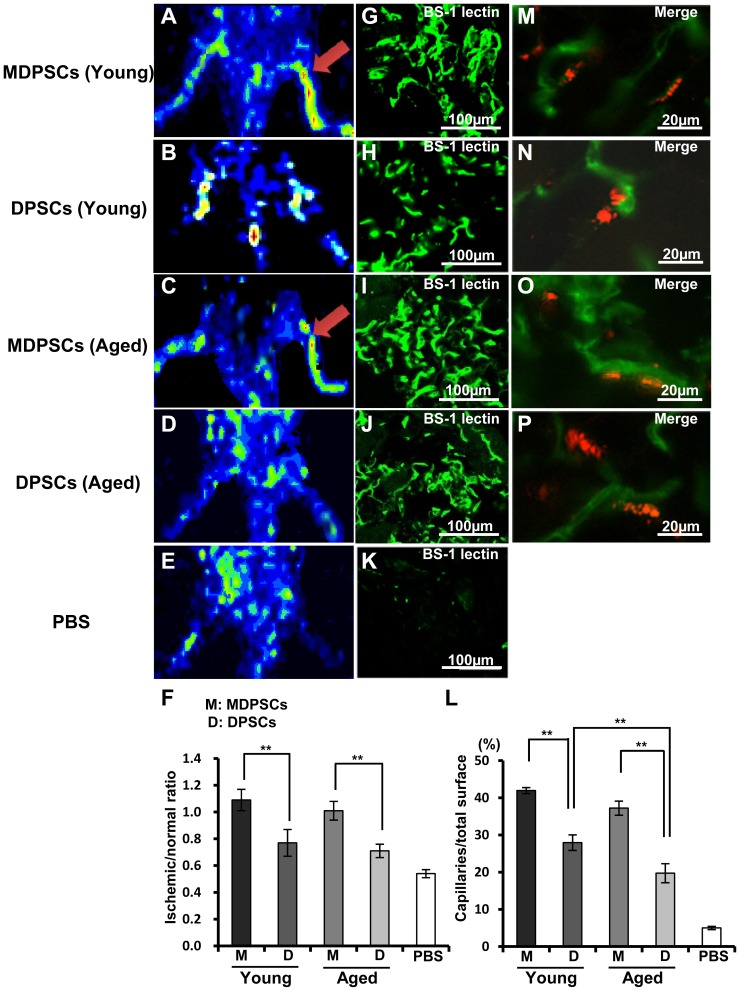
Figure 5. Neovascularization in the ischemic hindlimb 14 days after transplantation of aged MDPSCs.
Neovascularization in the ischemic hindlimb 14 days after transplantation of aged MDPSCs compared with those of aged DPSCs, young MDPSCs and young DPSCs. (A–E) Laser Doppler imaging. Accelerated blood flow (arrows). (F) Quantification of blood flow in the ischemic versus normal limbs obtained from four mice in each group. **p<0.01. (G–K) Immunostaining of Fluorescein Griffonia (Bandeiraea) Simplicifolia Lectin 1/fluorescein-galanthus nivalis (snowdrop) lectin (BS-1 lectin) in the ischemic hindlimb. (L) Quantification and statistical analysis of the capillary density in the ischemic region using serial sections. Data are expressed as means ± SD of 4 determinations. **p<0.01. The experiments were repeated three times (3 lots), and one representative experiment is presented. (M–P) Localization of DiI-labeled transplanted cells and newly formed capillaries stained by BS-1 lectin.

