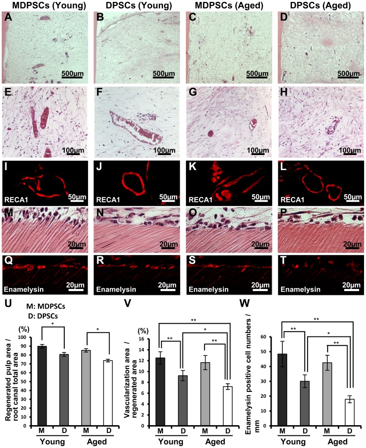
Figure 6. Regeneration of pulp tissue after ectopic tooth transplantation in severe combined immunodeficiency (SCID) mice.
Aged MDPSCs, aged DPSCs, young MDPSCs and young DPSCs were injected into the emptied root canals. (A–L) Hematoxylin and Eosin (HE) staining. (I–L) Immunostaining with RECA1. (M–P) Odontoblast-like cells lining to the dentinal wall. (Q–T) In situ hybridization analysis with enamelysin. (U) Ratio of the regenerated area to the root canal area. Data are expressed as means ± SD of four determinations. *p<0.05. (V) Ratio of the vascularization area to the regenerated area. Data are expressed as means ± SD of four determinations. *p<0.05, **p<0.01. (W) Enamelysin positive cell number of the dentinal wall. Data are expressed as means ± SD of four determinations. *p<0.05, **p<0.01.

