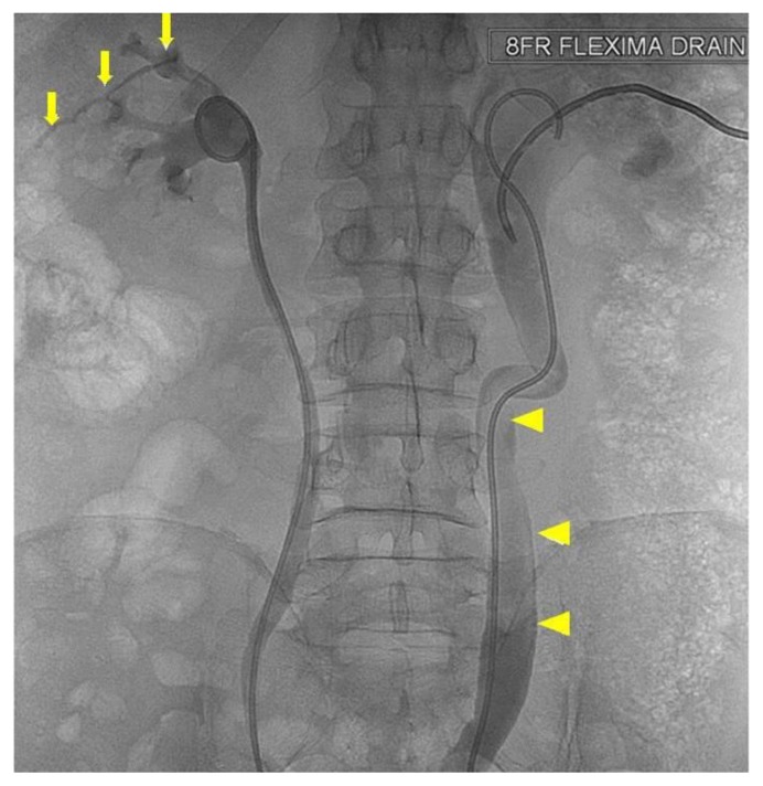
Figure 3.
64 year old male with an iatrogenic left urinothorax. Nephrostogram after initial antegrade stenting (patient prone). No covering nephrostomy has been placed after stenting on the left. Contrast can be seen extravasating from the left superior calyx (arrows) and a large clot lies within the lumen of the dilated right ureter (arrowheads). Spot fluoroscopic image was obtained after the anterograde injection of non-ionic water-soluble contrast material.

