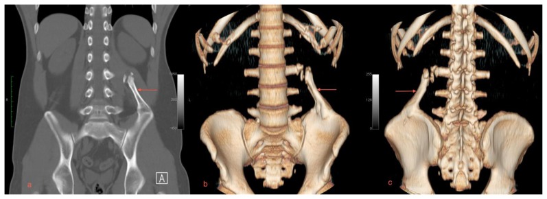
Figure 2.
A 29 year old male with myositis ossificans of the quadratus lumborum muscle. Protocol: Non-contrast enhanced CT, Siemens Somatom. Images were acquired kVp 120, mAs 51, helical acquisition, pitch 0.8, the original image collimation was 1.2 mm, the final images were reformatted to 5 mm thickness.
a) A coronal plane MIP reformat demonstrates the mature bone arising from the left iliac crest projecting toward the upper lumbar spine (arrow).
b) An anteriorly oriented 3D volume rendered CT image shows the relationship of the quadratus lumborum lesion with the L3 transverse process (arrow).
c) A posteriorly oriented 3D volume rendered CT image shows the relationship of the quadratus lumborum lesion with the L3 transverse process (arrow).

