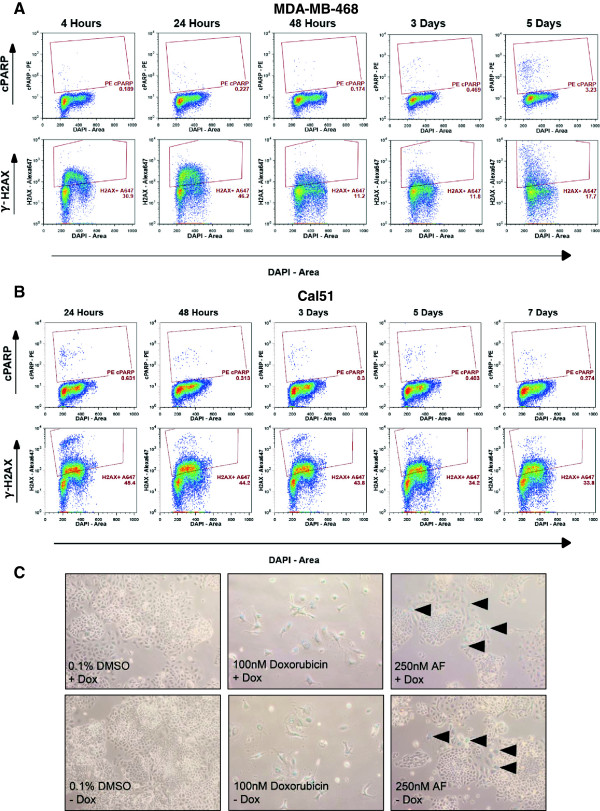
Figure 6.

AF induces DNA damage in MDA-MB-468 and Cal51, apoptosis in MDA-MB-468, and cellular senescence in Cal51shAhR. The presence of γ-H2AX and cleaved PARP (cPARP) was evaluated using fluorescent antibody based flow cytometry in MDA-MB-468 (A) and Cal51 (B) cells. Cells were treated with 25nM AF (MDA-MB-468) or 250nM AF (Cal51) for the indicated periods of time and stained with the appropriate fluorescent antibody, per protocol requirements. Raw flow cytometry data is shown. Samples were run on a BD LSR II flow cytometer. Appropriate fluorescent minus one samples were used to gate and analyze sample data. (C) The presence of AF-induced cellular senescence in Cal51shAhR was examined by staining for senescence-associated β-Galactosidase. Cal51shAhR cells were pretreated with 750 ng/mL Dox or an equivalent amount of vehicle to induce AhR knockdown. Cells were then treated with 0.1% DMSO or 250nM AF (nine days) or 100nM of a known inducer of senescence, doxorubicin (five days), with or without co-treatment with 750 ng/mL Dox. Cells were then fixed and stained with an X-Gal-containing staining buffer. Images taken at 10x are shown.
