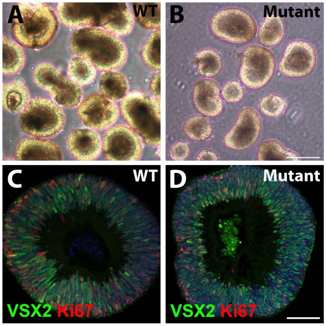
Figure 2. WT and (R200Q)VSX2 mutant hiPSC lines produce optic vesicle-like structures (hiPSC-OVs), allowing purification of VSX2+ neural retina progenitor cells (NRPCs).
(A, B) Representative micrographs of WT (A) and mutant (B) hiPSC-OVs isolated at day 20 of differentiation. At this stage, hiPSC-OVs are morphologically indistinguishable between WT and mutant cultures. Scale bar in panel B = 250 μm (also applies to panel A). (C, D) Both WT (C) and mutant (D) hiPSC-OVs were comprised of VSX2+ NRPCs that were proliferative, as shown by Ki-67 expression. Scale bar in panel D = 50 μm (also applies to panel C).

