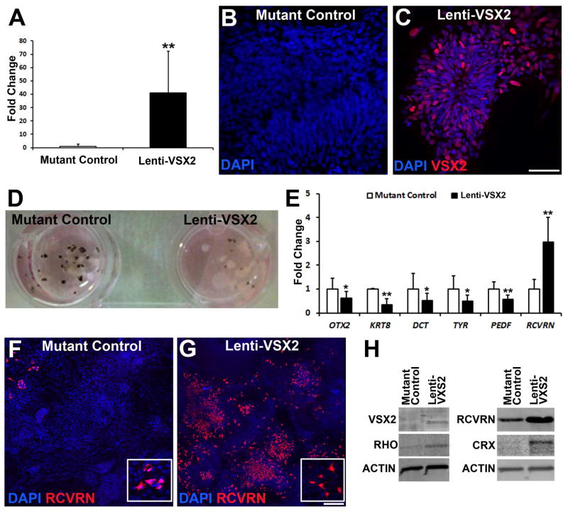
Figure 5. Exogenous expression of WT VSX2 reduces RPE production and enhances photoreceptor development in (R200Q)VSX2 hiPSC-OVs.
(A) Overall VSX2 expression was significantly increased in day 70 mutant hiPSC-OVs after transduction at day 14 with a WT VSX2-expressing lentiviral construct (Lenti-VSX2). Mutant control hiPSC-OVs were transduced at day 14 with a lenti-GFP construct. (B,C) At day 80, VSX2 was absent in lenti-GFP infected mutant hiPSC-OV controls (B), but remained expressed in mutant hiPSC-OV cells infected with lenti-WT VSX2 (C). Scale bar = 50 μm in panel C (also applies to panel B). (D) By day 70, exogenous expression of WT VSX2 in mutant hiPSC-OVs resulted in reduced pigmentation compared to lenti-GFP infected mutant hiPSC-OV controls. (E) qRT-PCR analysis at day 70 demonstrated reduced expression of characteristic RPE genes and increased expression of the photoreceptor gene RCVRN in lenti-WT infected vs. lenti-GFP infected mutant hiPSC-OVs. (F) At day 80, few RCVRN+ photoreceptors were present in lenti-GFP infected mutant hiPSC-OV control cultures. (G) Exogenous expression of WT VSX2 rescued RCVRN expression in mutant hiPSC-OVs. Insets in panels F and G demonstrate the cytoplasmic nature of RCVRN expression. Scale bar in panel G = 100 μm (also applies to panel F). (H) Western blot analysis at day 100 revealed increased expression of the photoreceptor genes RHO, CRX, and RCVRN in lenti-WT VSX2 infected mutant hiPSC-OVs when compared to lenti-GFP infected mutant hiPSC-OV control cultures. *p < 0.05, ** p < 0.01.

