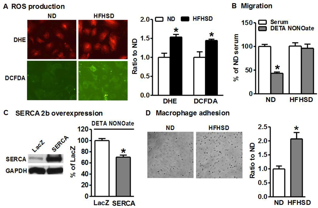
Figure 3. Smooth muscle cells from HFHSD-fed mice have increased ROS and macrophage adhesion, and impaired NO-inhibited migration.
A. ROS production in aortic SMCs is increased after 16 week HFHSD. Quantitation of fluorescence intensities in graph. *p < 0.05 vs. ND, n=3. B. NO donor fails to inhibit migration of cultured aortic SMCs isolated from HFHSD-fed mice. *p < 0.05 vs. serum, n=12. C. Overexpression of SERCA2b restores the inhibition of cell migration by NO donor in HFHSD SMCs. *p < 0.05 vs. LacZ (vector control), n=5. D. HFHSD enhances macrophage adhesion to cultured SMCs, quantified in graph. *p < 0.05 vs. ND, n=6.

