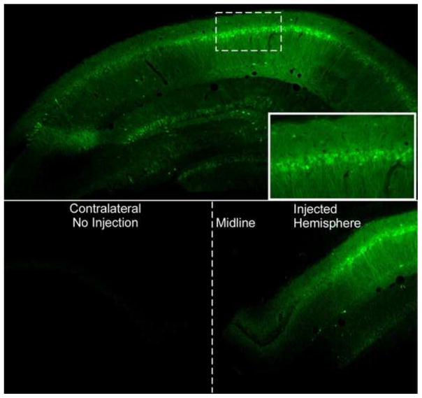
Figure 4. Green Flourescent Protein (GFP) expression in the hippocampus.
A: GFP expression in the hippocampus following infusion with serotype 2.1 AAV expressing GFP. Note the high level of expression in the pyramidal cells corresponding to the CA regions. Solid line box inset is shows close-up of pyramidal cells expressing GFP from the CA1 region (Dotted box). B: Note that GFP expression is only found in the hippocampus that was injected during a unilateral injection. The contralateral side within the same animal contained no cells expressing GFP. Midline of the hemispheres is denoted with a dotted line.

